
Allergen Testing - CAM 051
Description
Allergic disease is characterized by inappropriate or exaggerated rated immune reactions to foreign antigens (allergens) that are generally innocuous to most people, but when introduced into a genetically-predisposed individual, elicit a hypersensitivity reaction.1 Hypersensitivity reactions can be classified into four types, two of which are associated with allergy, type I immediate immunoglobulin E (IgE) reactions and type IV T cell–mediated reactions.2 Type I reactions involve the formation of IgE antibodies specific to the allergen. When the subject is re-exposed to that allergen, the allergen binds multiple IgE molecules, resulting in the release of an array of inflammatory mediators, including histamines, that precipitate the symptoms of allergic disease.1
Allergen testing in serum is designed to detect the presence of allergen-specific IgE. A positive test for allergen-specific IgE confirms the presence of the antibody only. Actual reactivity must be determined by history or supervised challenge.3 Several diagnostic procedures have been developed to elicit and assess hypersensitivity reactions including epicutaneous, intradermal, patch, bronchial, exercise, and ingestion challenge tests.4
Regulatory Status
The FDA has cleared 44 assays for total IgE as of July 13, 2021 (FDA, 2021). Additionally, many labs have developed specific tests that they must validate and perform in house. These laboratory-developed tests (LDTs) are regulated by the Centers for Medicare & Medicaid Services (CMS) as high-complexity tests under the Clinical Laboratory Improvement Amendments of 1988 (CLIA ’88). FDA clearance or approval is not currently required for clinical use.
Allergy testing for CMS plans may be covered when Medicare coverage criteria are met. Refer to the Medicare Claims Processing Manual Chapter 12 — Physicians/Nonphysician Practitioners for details. In addition, Local coverage determinations (LCDs)/local coverage articles (LCAs) are available for reference. Compliance with these policies is mandatory where applicable. Find these LCDs/LCAs at https://www.cms.gov/medicare-coverage-database/new-search/search.aspx (CMS, 2021).
Policy:
Application of coverage criteria is dependent upon an individual’s benefit coverage at the time of the request.
- When limited to allergens chosen for testing based on an individual’s history, physical examination, and environment, specific IgE in-vitro allergy testing (up to 20 allergen specific antibodies per year) is considered MEDICALLY NECESSARY.
- In-vitro testing for total serum IgE is considered MEDICALLY NECESSARY in any of the following situations:
- For individuals with moderate to severe asthma.
- For individuals with signs or symptoms of allergic bronchopulmonary aspergillosis.
- To monitor for allergy resolution in children and adolescents with an initial positive food allergen result(s), annual re-testing for the same food allergen(s) is considered MEDICALLY NECESSARY.
- When performed on the same day as skin prick testing, specific IgE in-vitro allergy testing is considered NOT MEDICALLY NECESSARY.
- In the absence of a new clinical presentation, routine re-testing for allergies to the same allergens (except where specified above) is considered NOT MEDICALLY NECESSARY..
- The antigen leukocyte antibody test (ALCAT) is considered NOT MEDICALLY NECESSARY..
The following does not meet coverage criteria due to a lack of available published scientific literature confirming that the test(s) is/are required and beneficial for the diagnosis and treatment of an individual’s illness.
- For individuals with signs or symptoms of allergies, basophil activation flow cytometry testing and in-vitro testing of IgG, IgA, IgM, and/or IgD is considered NOT MEDICALLY NECESSARY.
- In-vitro allergen testing using bead-based epitope assays (e.g., VeriMAP Peanut Dx) is considered NOT MEDICALLY NECESSARY..
- For all situations, in-vitro testing using qualitative specific IgE multi-allergen screen that does not identify a specific allergen is considered NOT MEDICALLY NECESSARY.
Policy Guidelines
Skin testing is contraindicated in the following situations:
- Patients who have certain skin conditions (e.g., dermatographism, urticaria, cutaneous mastocytosis, atopic dermatitis, severe diffuse psoriasis)
- Patient who are taking medications that may interfere with the treatment of anaphylaxis (e.g., Beta-blockers and Angiotensin Converting Enzyme inhibitors) or may impair skin test sensitivity (e.g., tricyclic antidepressants, antihistamines)
- Patients who are at high risk to testing (e.g., poorly controlled asthma, clinical history of severe reaction to minute amounts of allergen, cardiac arrhythmia, unstable angina)
- Patients who have experienced an anaphylactic event within the past one month
- Uncooperative patients (e.g., small children, individuals with mental or physical impairments)
Table of Terminology
| Term |
Definition |
| AAAAI |
The American Academy of Allergy, Asthma, and Immunology |
| AAAI |
Academy of Allergy, Asthma, and Immunology |
| AAFP |
American Academy of Family Physicians |
| AAP |
American Academy of Pediatrics |
| ACAAI |
American College of Allergy, Asthma, and Immunology |
| ACD |
Allergic contact dermatitis |
| ACR |
American cockroach |
| AIT |
Allergen immunotherapy |
| ALCAT |
The Antigen Leukocyte Antibody Test |
| AR |
Allergic rhinitis |
| Arah2-sIgE |
Arachis hypogaea 2-specific immunoglobulin E |
| ARIA |
Allergic Rhinitis and its Impact on Asthma |
| ASCIA |
Australian Society of Clinical Immunology and Allergy |
| ASIgE |
Allergen specific Immunoglobulin E |
| ATP |
Atopy patch test |
| AUC |
Area under the curve |
| BAT |
Basophil activation flow cytometry testing |
| BAT |
Basophil activation test |
| BBEA |
Peanut bead-based epitope assay |
| CAA |
Current allergic asthma |
| CAR |
Current allergic rhinitis |
| CBS |
Consensus based statements |
| CD4+ |
Cluster of differentiation 4 |
| CLIA ’88 |
Clinical Laboratory Improvement Amendments of 1988 |
| CLSI |
Clinical and Laboratory Standards Institute |
| CMS |
Centers for Medicare & Medicaid Services |
| CRS |
Chronic rhinosinusitis |
| CW |
Choosing wisely |
| EAACI |
European Academy of Allergy and Clinical Immunology |
| EDTA |
Ethylenediaminetetraacetic Acid |
| EP |
Expert panel |
| FA |
Food allergy |
| FcεRI |
High-affinity IgE receptor |
| FDA |
The Food and Drug Administration |
| FEV1 |
Forced expiratory volume in 1 second |
| GA2LEN |
Global Allergy and Asthma European Network |
| H1 |
H-1 receptor antagonists |
| H2 |
H-2 receptor antagonists |
| IgA |
Immunoglobulin A |
| IgD |
Immunoglobulin D |
| IgE |
Immunoglobulin E |
| IgE-FAB |
Immunoglobulin E- fragment antigen-binding region |
| IgG |
Immunoglobulin G |
| IgG4 |
Immunoglobulin A |
| IgM |
Immunoglobulin A |
| JTFPP |
Joint Task Force on Practice Parameters |
| LCD |
Local coverage determination |
| LDT |
Laboratory-developed test |
| LEAP |
Learning early about peanut allergy |
| MBB |
Mucosal brush biopsies |
| MFI |
Median fluorescence intensity |
| NASEM |
The National Academies of Science, Engineering and Medicine |
| NIAID |
National Institute of Allergy and Infectious Diseases |
| NICE |
National Institute for Health and Care Excellence |
| NPV |
Negative predictive value |
| NSAID |
Nonsteroidal anti-inflammatory drug |
| OFC |
Oral food challenge |
| PAMD@ |
Precision allergy molecular diagnostic applications |
| PPV |
Positive predictive value |
| RARS |
Recurrent acute rhinosinusitis |
| SAR |
Seasonal allergic rhinitis |
| sIgE |
Specific immunoglobulin E |
| s-IgE |
Specific immunoglobulin E |
| SPT |
Skin prick tests |
| SSRI |
Selective serotonin reuptake inhibitor |
| ST |
Skin test |
| Th2 |
T helper type 2 |
| tIgE |
Total immunoglobulin E |
| WAO |
World Allergy Organization |
Rationale
Allergies affect about 30 percent of Americans.5 The incidence of allergic disease is increasing6 and is estimated to result in over $17 billion in health care costs and 200,000 emergency department visits annually.7
A majority of environmental, food, and medication allergies with clinical significance are type I immunoglobulin E (IgE)-mediated allergies.3 Diagnosis of an IgE-mediated allergy involves identification of the allergen, demonstration of IgE-specific to that allergen, and confirmation that symptoms occur when the patient is exposed to the allergen. The IgE response to an allergen can be assessed using skin or serum testing. Patch testing is preferred for delayed T cell–mediated response.2,8
Allergic diseases, respiratory infections, and autoimmune conditions have similar clinical presentations and self-reported symptoms have a relatively low positive predictive value (PPV).9 Thus, laboratory allergy and immunologic testing are useful in clarifying diagnosis and guiding treatment when the frequency, duration, and sequelae of upper respiratory infections exceed the norm or when rhinosinusitis or asthma symptoms persist despite treatment.10 Total IgE can be useful in diagnosing asthma and differentiating from other conditions such as allergic bronchopulmonary aspergillosis (ABPA) which is characterized by signs and symptoms that may include fever, hemoptysis, coughing up of mucus plugs, shortness of breath, chest or joint pain, headaches, skin lesions, wheezing, worsening asthma, unintentional weight loss and/or fatigue.11 Allergy testing is also useful in identifying causative allergen in atopic dermatitis (eczema), contact dermatitis, urticaria, angioedema, and food or drug allergies. Knowing the causal allergen helps provide clinically relevant information for avoidance and treatment.2
Skin testing
Skin testing is the most rapid, sensitive, and cost-effective testing modality for the detection of IgE-mediated disease. The procedure lasts less than an hour with minimal patient discomfort. There are several published practice parameters for allergen skin testing.2-4
Serum IgE
Immunoglobulin E (IgE) is one of five immunoglobulins and the one primarily involved in allergic disease. At the cellular level, the allergic response starts with “atopy,” a genetic predisposition to produce specific IgE after exposure to allergens. CD4+ helper T cells are predisposed to the “T helper type 2” (Th2) response, which causes the Th2 cells to secrete large amounts of interleukins 4 and 13, which then promotes production of the allergen-specific IgE. From there, the allergen-specific IgE binds to high-affinity receptors on mast cells and basophils. At this point, if the relevant allergen is ingested in large enough amounts, the IgE molecules may cluster (cross-linking). This cross-linking causes the mast cells and basophils to release chemical and protein mediators, resulting in the characteristic allergic response.12
Immunoassays measuring both total IgE and allergen-specific IgE in serum and other bodily fluids have been developed. Specific IgE immunoassays do not require patient cooperation, are not limited in patients with skin disease, are not blocked by antihistamines, and pose no risk of adverse reactions.2,4,12,13 Total IgE is usually unrelated to IgE levels for a specific allergen but may be useful in other conditions, such as asthma.12
Other testing
Patch testing is the gold standard for identification of a contact allergen.14,15 Although occlusive patch testing is the most common technique, open, prophetic (provocative), repeated insult, photopatch, and atopy patch tests are also available if special situations indicate their use.4
Cellular activation assays measuring the release of histamine from basophils16,17 or mast cells18 as diagnostic or prognostic indicators of allergy have been the subject of intense research. Basophil and eosinophilic reactivity tests have been found to be associated with food-induced allergic responses and have been shown in current research to be modified over time during immunotherapy.9 In particular, the basophil activation test (BAT) has emerged as having superior specificity and comparable sensitivity to diagnose food allergies when compared with skin prick test and specific IgE.19 Histamine release from leukocytes of allergic persons is an excellent in-vitro correlate of allergy; however, it is currently still considered a research test by the Academy of Allergy, Asthma, and Immunology.4 In-vitro tests can be used to confirm a negative skin test. Skin testing has a higher sensitivity and is less expensive compared to in-vitro. In-vitro could also be useful if an individual has certain skin conditions that could produce false-positive results.3
Basophil activation flow cytometry testing (BAT) has the potential to be a useful tool for measuring hypersensitivity to allergens, especially for patients who are not suitable for skin testing due to skin status or prior severe reactions since it is an ex vivo, flow cytometry-based assay. BAT, for use as standard clinical practice, is currently limited by its lack of standardization in methodology as well as between systems used. A study by Depince-Berger, et al. (2017) has proposed standardization between systems and instruments using whole blood-ethylenediaminetetraacetic acid (EDTA) samples with instrumentation standardization. “BAT would strongly benefit from easy implementation [EDTA, one step stimulation/labeling, wash, full sample analysis over time parameter, B cell relative basophil count] and standardization of instrument settings on MFI [median fluorescence intensity] targets whatever system or instrument is used.”20 Hemmings, et al. (2018) note that standardization, quality assurance, and clinical validation will facilitate the transition of the BAT from research to clinical practice.
Proprietary testing
The Antigen Leukocyte Antibody Test (ALCAT) is another test available for the assessment of allergens. ALCAT measures food/immune reactions through stimulation of leukocytes. The immunological reactions to this stimulation are intended to identify sensitivities regardless of pathway as antibodies do not necessarily need to be involved. Cell Science Systems suggests individuals with a variety of disorders (such as gastrointestinal, neurological, et al.) to take this test.22 Although the ALCAT machine is FDA registered and there are a few papers published, results are not reproducible when subject to rigorous testing and do not correlate with clinical evidence of allergy.23-25
Panels encompassing a large number of analytes are also offered by labs. For example, Genova Diagnostics offers a blood test for IgG and IgE antibodies for 87 different foods. Genova also offers several variations on this test, such as “Vegetarian” (21 foods), “Spices” (23 spices), “Molds” (15 molds), “Inhalants” (16 inhalants specific to 18 North American geographic regions), and “Celiac profile” (Total IgA, tTG IgA & IgG, DGP IgA & IgG, EMA IgA).26
Spiriplex offers a microarray-style panel for allergen testing, called “Allergenex.” This test contains many purified allergen proteins to which a patient’s blood sample can bind. This binding creates a quantifiable signal that allows the user to identify the number of IgE antibodies present, and therefore provide a picture of allergy. Spiriplex offers a test for 28 common food allergens, a test for 40 inhalant allergens, and 68 combined food and inhalant allergens.27
The VeriMAP Peanut Dx and the VeriMAP™ Peanut Sensitivity are both peanut allergen specific bead-based epitope assays manufactured by AllerGenis LLC. According to Allergenis, VeriMAP™ has a “95% positive predictive value and can reduce overdiagnosis and anxiety by minimizing false positives.”28 This is an emerging technology and additional peer-reviewed literature establishing the analytical validity, clinical validity, and clinical utility of such testing will be further required.
Analytical Validity
Variables that can influence the wheal size when performing skin prick tests (SPT) include multiple operators, extract concentrations and quality, skin test devices, time of day, location on the skin, and the measuring of results.29,30
In 2006, Oppenheimer and Nelson evaluated variability and analytical validity of skin testing. A questionnaire was sent to all physician and fellow members of the American College of Allergy, Asthma and Immunology who were currently practicing in the United States. The objective of this questionnaire was to determine the diversity of skin testing practices among allergists. The results showed great variability among physicians. In particular, “The average number of skin prick tests performed ranged from 5.09 (grasses) to 10.9 (trees), whereas the average number of intradermal tests performed ranged from 2.03 (grasses) to 5.6 (perennial). The allergen extract concentrations used for intradermal testing varied widely. Expressed as a dilution of the concentrated extracts, 20.8% use 1:100 dilutions, 10.3% use 1:500 dilutions, and 59.4% use 1:1,000 dilutions. Significant variability also occurred regarding devices and the technique with which the devices were used. Most clinicians (92.1%) used the most concentrated extract available for skin prick testing. For reporting the results of skin testing, 53.8% used a 0 to 4+ scale, and only 28.3% measured orthogonal diameters. Of those using a 0 to 4+ scale, two thirds related the results to the size of the histamine control.”31 The results from this survey emphasize potential areas of improvement for allergists regarding skin test use and data.
The Clinical and Laboratory Standards Institute (CLSI) has evaluated the analytical validity of serum IgE measurements and found that “Clinical/diagnostic sensitivity and specificity of IgE antibody assays cannot be accurately determined due to the absence of definitive gold standard methods for defining allergic disease. Total and allergen-specific IgE analyses achieve among the highest analytical performance of any antibody assay by following consensus procedures in CLSI-ILA20-A3.”32
Knight, et al. (2018) “examined the qualitative concordance between SPT and sIgE as measured on the HYTEC™288 platform for 10 commonly encountered inhalant allergens”; a total of 232 subjects were included. Overall concordance between SPT and sIgE was >70% for all allergens tested. Sensitivity ranged from 25% to 95% depending on the allergen, while specificity was significantly higher for all allergens (78-97%). Negative predictive value (NPV) was >85% for all allergens tested, while PPV was more variable, ranging from 22% to 88%. The authors noted that “these results are similar to findings in other studies comparing SPT with sIgE.”33
Carlsson, et al. (2015) examined the inter- and intra- variability of IgE and IgE receptor expression in the blood of seasonal allergic rhinitis (SAR) subjects. Thirty-two patients with SAR were included; the high-affinity IgE receptor, also known as FcεRI, and the low affinity receptor, also known as CD23, were measured. The authors found that “FcεRI expression on basophils and CD23 expression on B cells showed low intrasubject variability both in and out of the pollen season,” although there was a small seasonal difference with lower total IgE levels and FcεRI expression during the pollen season.34
Siroux, et al. (2017) explored the effect of allergen nature, route of exposure, and dose of exposure on IgE and IgG responses. A total of 340 patients (170 with asthma, 170 without) were included, and IgE/IgG responses to 47 inhalant and food allergens were analyzed and compared between five French regions according to route of allergen exposure (inhaled or food). “Ubiquitous” allergens (grass, olive/ash pollen, house dust mites) did not show marked difference in specific IgE level between regions. For region-specific allergens (ragweed, birch, cypress), IgE sensitization was associated with regional pollen exposure. Airborne allergens cross-reacting with food allergens led to frequent IgG recognition. The authors concluded that “the variability in allergen-specific IgE and IgG frequencies depends on exposure, route of exposure, and overall immunogenicity of the allergen. Allergen contact by the oral route might preferentially induce IgG responses.”35
Sookrung, et al. (2019) measured the agreement of a SPT and serum-specific IgE test to Periplaneta americana (American cockroach, ACR) allergies. ACR-extract was used, and sera was obtained from 66 individuals clinically diagnosed with chronic allergic rhinitis. Of the 66 samples, 46 were positive and 20 negative after a SPT to ACR-extract. Serum IgE levels were then measured by a commercial test kit. The authors note that of the SPT positive cases to ACR-extract, only 32.6% were also positive for serum IgE, indicating low concordance between the two testing methods.36
He and Reisacher (2019) measured the sensitivity, specificity, and predictive value of oral mucosal brush biopsies (MBB) as a new diagnostic test for peanut allergies. Twenty individuals participated in this study; each participant underwent oral MBB and serum testing for peanut IgE. The authors note that “At 0.12 kU/L, the sensitivity of oral MBB testing was 80% and the specificity was 85%, whereas at 1.0 kU/L, the sensitivity of sIgE testing was 50% and the specificity was 100%. From the ROC curves, the areas under the ROC curve (AUC) for oral MBB and sIgE were 0.91 (p < 0.001) and 0.74 (p = 0.007), respectively. Combination testing further increased both sensitivity and accuracy over oral MBB alone.”37 These results are promising for oral MBB, although more research needs to be completed.
Clinical Utility and Validity
In 1998, Tschopp, et al. (1998) compared three diagnostic tests for atopic diseases. Total serum IgE, Phadiatop, and the SPT were compared for 8329 individuals. Current allergic asthma (CAA) and current allergic rhinitis (CAR) were the conditions studied. The prevalence of CAA was 1.8% and prevalence for CAR was 16.3%. The prevalence of positive tests was 29%, 23%, and 23% for Phadiatop, SPT, and IgE, respectively. The results were as follows: “To diagnose current allergic asthma (CAA) and current allergic rhinitis (CAR), the sensitivity of Phadiatop was significantly higher than that of SPT (72.5% vs 65.4%, 77.1% vs 68.4% respectively) and IgE (72.5% vs 56.9%, 77.1% vs 43.9%, respectively). The sensitivity of SPT was significantly higher (68.4% vs 43.9%) than that of IgE to diagnose CAR. When CAA and CAR were excluded, the SPT specificity was significantly higher than that of Phadiatop (77.8% vs 71.9% and 85.9% vs 80.5%, respectively): when CAR was excluded, SPT was significantly higher than IgE (85.9 vs 81.4%). SPT had significantly the best positive predictive value for CAA (5.2% for SPT vs 4.6% for both IgE and Phadiatop) and CAR (48.7% for SPT vs 43.5% for Phadiatop and 31.6% for IgE). The three markers of atopy had roughly the same negative predictive value (NPV) for CAA, but IgE had a significantly lower NPV for CAR than SPT and Phadiatop (88.1% vs 93.3% and 94.7%, respectively). The diagnostic efficiency of SPT was significantly higher than that of Phadiatop (83.1% vs 79.9% and 77.6 vs 71.9%, respectively) to diagnose CAR and CAA. IgE and SPT had equal efficiency (77.6%), which was significantly higher than that of Phadiatop, to diagnose CAA (71.9%).”38 The authors concluded that “SPT have the best positive predictive value and the best efficiency to diagnose respiratory atopic diseases. Furthermore, SPT give information on sensitivity to individual allergens and should therefore be used primarily by clinicians to assess respiratory allergic diseases.”38
A retrospective analysis included patients who had been prick tested to “establish whether an incomplete diagnosis would have been reached if patch testing had been omitted.” The authors observed that if “investigation of allergic skin disease is undertaken by a non‐dermatologist, it is unlikely that patch testing will be performed.” A total of 330 patients had been prick tested in the time period specified. Sixty-eight patients had positive reactions on prick testing, and 36 of those had positive patch tests. Of the 262 patients who had negative prick tests, 121 had positive patch tests (46.1%) of current relevance to patient history in 92 subjects (35.1%). The authors concluded that “omission of patch testing from the investigation of allergic skin disease, even when contact urticaria may be the sole suspected diagnosis, would result in the frequent missed diagnosis of contact allergy.”39
In 2014, a meta-analysis examined the clinical validity of SPT and IgE measurement for food allergy. Twenty-four studies consisting of 2831 participants were included. The results were as follows: “For cows' milk allergy, the pooled sensitivities were 88% (SPT), and 87% (IgE) and specificities were 68% and 48%. For egg, pooled sensitivities were 92% and 93% and specificities were 58% and 49% for SPT and specific IgE. For wheat, pooled sensitivities were 73% and 83% and specificities were 73% and 43% for SPT and sIgE. For soy, pooled sensitivities were 55% and 83% and specificities were 68% and 38% for SPT and sIgE. For peanut, pooled sensitivities were 95% and 96%, and specificities were 61% and 59% for SPT and sIgE.”40
Klemans, et al. (2015) examined the diagnostic accuracy of using sIgE to peanut components to improve sensitivity and specificity of peanut allergen testing. Twenty-two studies were included. The authors found that “sIgE to Ara h 2 [a peanut component] showed the best diagnostic accuracy of all diagnostic tests to diagnose peanut allergy. Compared to the currently used SPT and sIgE to peanut extract, sIgE to Ara h 2 was superior in diagnosing peanut allergy.”41 The authors also found that the worst accuracy was observed to be sIgE to Ara8 and Ara9. The authors concluded that “sIgE to Ara 2 should replace SPT and sIgE to peanut extract in daily clinical practice.”41
Caglayan Sozmen, et al. (2015) examined the diagnostic accuracy of using the patch test to avoid oral food challenge (OFC). They found that in 243 children that underwent OFC to suspected food, clinically relevant food allergies were seen in 40 (65%) children to egg and in 22 (35%) to cow's milk. The sensitivity of SPT for both milk and egg were 92%, specificity 91%, PPV 35%, and NPV 93%. Sensitivity, specificity, PPV, and NPV of atopy patch test for both milk and egg were 21%, 73%, 20%, and 74%, respectively.
Santos, et al. (2014) studied the performance of BAT as a diagnostic marker for peanut allergy. Forty-three peanut-allergic children, 36 peanut-sensitized but tolerant children, and 25 non–peanut-sensitized non-allergic children underwent SPT, sIgE, and BAT. The authors found that BAT in peanut-allergic children showed a peanut dose-dependent upregulation of CD63 and CD203c while there was no significant response in the other two cohorts. BAT optimal diagnostic cutoffs showed 97% accuracy, 95% PPV, and 98% NPV. BAT allowed reduction of required OFCs by two-thirds. BAT proved particularly useful in cases in which specialists could not accurately diagnose peanut allergy with SPT and sIgE to peanut and to Arah2. Using a two-step diagnostic approach in which BAT was performed only after equivocal SPT or Arah2-sIgE, BAT had a major effect (97% reduction) on the number of OFCs required.
Santos, et al. (2015) also studied the utility of BAT to predict the severity and reactivity to peanut during OFCs. They found that “Of the 124 children submitted to OFCs to peanut, 52 reacted with clinical symptoms that ranged from mild oral symptoms to anaphylaxis. Severe reactions occurred in 41% of cases, and 57% reacted to 0.1 g or less of peanut protein. The ratio of the percentage of CD63(+) basophils after stimulation with peanut and after stimulation with anti-IgE (CD63 peanut/anti-IgE) was independently associated with severity, whereas the basophil allergen threshold sensitivity CD-sens (1/EC₅₀ × 100, where EC₅₀ = half maximal effective concentration) value was independently associated with the threshold of allergic reactions to peanut during OFCs. Patients with CD63 peanut/anti-IgE levels of 1.3 or greater had an increased risk of severe reactions (relative risk, 3.4). Patients with a CD-sens value of 84 or greater had an increased risk of reacting to 0.1 g or less of peanut protein (relative risk, 1.9)”.44 The authors concluded that “Basophil reactivity is associated with severity, and basophil sensitivity is associated with the threshold of allergic reactions to peanut. CD63 peanut/anti-IgE and CD-sens values can be used to estimate the severity and threshold of allergic reactions during OFCs.”44
Davila, et al. (2015) explored the association between total IgE and severity of asthma. A total of 383 patients were included (129 mild, 82 moderate, and 172 severe). Serum IgE levels were noted to vary “markedly” (147% coefficient of variation). The authors did not find an association between total IgE and forced expiratory volume in one second (FEV1) or asthma severity; although, the severe subgroup had a higher percentage of patients with >400 IU/mL. Independent predictors of higher IgE were found to be younger age, sensitization to at least two allergens, male gender, and family history of asthma. The authors concluded that “we did not find a significant association between serum total IgE levels and asthma severity or airflow limitation, except for a higher percentage of patients with IgE > 400 IU/mL in the severe subgroup.”45
Tannert, et al. (2017) investigated the relevance of a positive skin test and positive IgE test to penicillin allergy. Twenty-five patients with positive results were given penicillin, and another 19 patients deemed allergic were included. However, only nine of the 25 patients given penicillin were challenge positive. Positive results from each test alone did not predict allergy. The authors concluded that “the best predictor for a clinically significant (IgE-mediated) penicillin allergy is a combination of a positive case history with simultaneous positive ST result and s-IgE or a positive challenge result.”46
Suárez-Fariñas, et al. (2021) investigated the validity of the peanut BBEA diagnostic test on 133 subjects as well as on 82 additional subjects from another study, forming a cohort for a paper titled, “Accurate and reproducible diagnosis of peanut allergy using epitope mapping.” The authors measured levels of IgE to epitopes evaluated against a threshold established prior to the study. The peanut BBEA diagnostic test diagnosed 93% of subjects accurately, with a sensitivity threshold of 92% and specificity of 94%. The positive predictive value (PPV) was 91%. The authors concluded that “the overall accuracy was found to be superior to existing diagnostic tests for peanut allergy including skin prick testing, peanut sIgE, and peanut component sIgE testing.”47
Kwong, et al. (2024) analyzed “aeroallergen testing data from the 2019 serum IgE tests for seasonal inhalant allergens through Quest Diagnostics database. Individuals with results for at least 1 of 31 seasonal allergens across 4 allergen classes (11 trees, 7 weeds, 5 grasses, and 8 molds) were analyzed. Of 88,042 individuals tested for ≥1 seasonal allergen, 1.5%, 1.8%, 1.3%, and 1.6% were tested for all trees, weeds, grasses, and molds, respectively. Of those tested for all allergens within a class, 40.4%, 38.6%, 29.5%, and 21.2% were sensitized to at least one tree, weed, grass, or mold allergen. Identification of ≥98% of sensitized patients within a class required 8 allergens for trees (mountain cedar, maple box elder, walnut, white ash, elm, birch, cottonwood, and hickory/pecan), 5 for weeds (common ragweed short, rough pigweed, English plantain, lamb's quarters/goosefoot, and Russian thistle), 3 for grasses (June/Kentucky blue grass, Johnson grass, and Bermuda grass), and 7 for molds (Alternaria alternata, Aspergillus fumigatus, Mucor racemosus, Epicoccum purpurascens, Penicillium notatum, Helminthosporium halodes, and Fusarium moniliforme)”. The authors concluded that a minimum of 23 antigens is required to optimally detect sensitization to four classes of seasonal allergens. The addition of these allergens to unique perennial allergens (cat, dog, mouse, cockroach, and 2 dust mite species) results in a comprehensive elucidation of inhalant allergen sensitization.48
The American Academy of Allergy, Asthma, and Immunology and the American College of Allergy, Asthma, and Immunology (ACAAI)
The AAAI and ACAAI published practice parameters in 2008 for allergy testing which noted that “for individual patients, the choice of test allergens is guided by the history and physical examination and the physician’s knowledge, training, and experience.”4 The guidelines recommended that “specific IgE immunoassays may be preferable to skin testing under special clinical conditions, such as widespread skin disease, patients receiving skin test suppressive therapy, uncooperative patients, or when the history suggests an unusually greater risk of anaphylaxis from skin testing.” They also note that for both skin testing and in-vitro specific IgE testing, “the allergens selected … should be determined based on the patient’s age, history, environment and living conditions (e.g., region of the country), occupation, and activities.” Also, “the best indicators in the selection of appropriate pollens for clinical use are extensive prevalence in the air and concurrent allergy symptoms during annually recurrent seasons when such pollens are expected to be present in the ambient air.”
The AAAAI state that symptoms including itchy eyes, nose or throat; nasal congestion, runny nose, sneezing, watery eyes, chest congestion, cough or wheezing, eczema, severe site reaction to insect stings, anaphylaxis, and reactions to foods warrant an allergy test. IgE skin testing is the preferred method to testing. Blood tests are used when a skin test is not considered safe or useful.49
The AAAAI and ACAAI guidelines also state, “as is the case with skin tests, a direct correlation cannot be assumed between the presence of specific IgE (sIgE) antibodies and clinical disease.” Additionally, “sensitivity and the positive predictive value of both prick/puncture and specific IgE tests generally tend to be higher among pollens, stable anaphylactogenic foods, house dust mite, certain epidermals, and fungi compared with venoms, drugs, and chemicals.”
With regards to total IgE testing, these groups indicate, “measurements of total serum IgE concentration are of modest clinical value when used as a screen for allergic disease or for predicting the risk of allergic disease.”
The AAAAI and ACAAI also note that “IgG and IgG subclass antibody tests for food allergy do not have clinical relevance, are not validated, lack sufficient quality control, and should not be performed.”
Regarding basophil activation assays, they state, “histamine and leukotriene release measurements from human basophils after incubation with allergen are valuable research tools for in vitro investigations of allergy.”4
Their practice parameter on drug allergy also states that “the basophil activation test is a recently described method of evaluating expression of CD63 on basophils after stimulation with an allergen. There are limited data using this method to evaluate patients with possible allergies to β-lactam antibiotics and nonsteroidal anti-inflammatory drugs (NSAIDs).”50
They also recommend, “because anaphylactic reactions cannot be distinguished from anaphylactoid, nonimmune occurrences, it has been recommended that plasma histamine, tryptase, and specific IgEs (if available) may be ordered at the time of reaction and skin tests be performed later.”50
In their 2014 practice parameter on food allergy they acknowledge: “Basophil and eosinophilic reactivity tests have been shown to be associated with food-induced allergic responses and have been shown in current research to be modified over time during immunotherapy.”9
Their 2014 practice parameter on rhinosinusitis also recommends to “perform an evaluation for specific IgE antibodies to airborne allergens in patients with RARS or CRS.” An updated practice parameter on rhinitis published in 2020 comments that local allergic rhinitis will often be associated with “negative skin prick tests (and intradermal tests, when performed) and absence of serum-specific IgE (sIgE) antibodies but a positive nasal allergen provocation test (NAPT) to aeroallergens.51 With respect to vasomotor rhinitis, the authors state that “laboratory tests, skin prick tests, and sIgE are helpful only to exclude AR [allergic rhinitis].”51
In this practice parameter, they also make the following summary concerning re-evaluation of food allergies in children and adolescents: “Summary Statement 11: Consider the natural course of allergies to specific foods when deciding on the frequency of food allergy follow-up evaluations, recognizing that allergies to certain foods (milk, egg, wheat, and soy) generally resolve more quickly in childhood than others (peanut, tree nuts, fish, and shellfish). These observations could support individualized follow-up (ie, roughly yearly re-evaluations of these allergies in childhood) with less frequent retesting if results remain particularly high (eg, >20-50 kUA/L). [Strength of recommendation: Moderate; C Evidence].”51
In their 2015 practice parameter on anaphylaxis,52 they recommend “skin tests and/or in vitro tests for specific IgE and challenge tests might be appropriate to help define the cause of the anaphylaxis.”
They also recommend against routinely obtaining total serum IgE levels for the diagnosis of food allergy, however, because of the low PPV of self-reported symptoms and lack of pathognomonic signs on physical examination, they recommend that the accurate diagnosis of IgE-mediated food allergy should be aided by laboratory allergy testing, including skin prick and/or serum IgE testing. The clinician should use specific IgE tests (SPTs, serum tests, or both) to foods as diagnostic tools; however, testing should be focused on foods suspected of provoking the reaction, and test results alone should not be considered diagnostic of food allergy. Moreover, “the diagnosis of food-induced anaphylaxis should be based on signs and symptoms in association with likely or known exposure to a food allergen”, as “events mimicking anaphylaxis also can occur after the ingestion of food.”52
The practice parameter on anaphylaxis was updated in 2023. New recommendations include “We suggest that immediate hypersensitivity skin testing (percutaneous and intradermal) and/or in vitro-specific IgE testing be performed, when available, to all potential pharmacologic and nonpharmacologic culprits used during the perioperative period. If testing is not possible, we suggest referral to another center or, if necessary, use of the most efficacious agents structurally dissimilar from the most likely culprit.” And, “We suggest that immediate hypersensitivity testing to suspected culprit (and alternative) agents be delayed after [perioperative anaphylaxis], unless repeat surgery cannot be postponed. If surgery with general anesthesia is needed sooner, then testing may be performed when needed.” Lastly, “We suggest that challenges be performed, when feasible, to all potential culprit agents to which skin and/or in vitro testing is negative, before or in conjunction with use of these agents for a future surgical procedure.”53
In a Choosing Wisely (CW) report, the AAAAI recommends against performing “unproven diagnostic tests, such as immunoglobulin G (lgG) testing or an indiscriminate battery of immunoglobulin E (IgE) tests, in the evaluation of allergy.”54
In another CW report, the AAAAI recommends against routine diagnostic testing in patients with chronic urticaria, stating that “skin or serum-specific IgE testing for inhalants or foods is not indicated, unless there is a clear history implicating an allergen as a provoking or perpetuating factor for urticaria.”54
The AAAAI also published a 2020 practice parameter update on peanut allergy diagnosis. The authors recommend in favor of diagnostic SPT or sIgE testing for peanut allergy in patients with physician-judged high pretest probability of peanut allergy. Testing is also recommended prior to an OFC for patients with moderate pretest probability of peanut allergy. Ara h 2 diagnostic testing is the suggested approach for patients presenting for evaluation of suspected peanut allergy for which a single diagnostic test is to be used, due to its superior diagnostic accuracy “by virtue of more optimal positive/negative likelihood ratios.” However, Ara h 2 is noted to have lower sensitivity than the skin prick or sIgE tests, so a clinician may use Ara h 2, SPT, or sIgE to confirm the diagnosis of peanut allergy in a patient with a high prior probability. The AAAAI recommends against “routine use of component testing in addition to either SPT or sIgE to whole peanut to increase diagnostic accuracy,” and against using the results of skin prick or sIgE to determine “the severity of an allergy phenotype or to predict the severity of a future reaction.”55 It is noteworthy that all the recommendations above were assigned “low” or “very low” degrees of evidence certainty.55
Joint Task Force on Practice Parameters (JTFPP)
In a practice parameter concerning contact dermatitis, the Joint Task Force on Practice Parameters—composed of the American Academy of Allergy, Asthma & Immunology (AAAAI), the American College of Allergy, Asthma & Immunology (ACAAI), and the Joint Council of Allergy, Asthma & Immunology—proposed this series of summary statements:
“Summary Statement 1: Consider ACD [allergic contact dermatitis] in the differential diagnosis of patients with chronic eczematous or noneczematous dermatitis. [Strength of Recommendation: Strong; C Evidence]
Summary Statement 2: In patients suspected of ACD, patch testing is the gold standard to confirm the diagnosis. [Strength of Recommendation: Strong; C Evidence]
Summary Statement 3: In addition to personal products used by a patient suspected of ACD, review the home and workplace for other sources of contact allergens. [Strength of Recommendation: Moderate; D Evidence] Summary Statement
Summary Statement 4: Evaluate patients for both irritant and allergic causes, especially in those presenting with hand dermatitis. [Strength of Recommendation: Strong; C Evidence]
Summary Statement 5: Allergic CD should be suspected and evaluated in the patient with both generalized and anatomically localized skin eruptions (such as the hands, face, eyelids) that come in contact with the substances in the environment. [Strength of Recommendation: Moderate; C Evidence]
Summary Statement 6: In a patient with a facial rash involving the periorbital areas (eg, eyelids), evaluate for ACD caused by components of cosmetics, such as fragrances, preservatives, and excipients, because these are common sensitizers of the facial skin. [Strength of Recommendation: Moderate; C Evidence]
Summary Statement 7: Evaluate patients presenting with lip dermatitis (cheilitis) and perioral dermatitis for both irritant and allergic causes of contact dermatitis. [Strength of Recommendation: Moderate; C Evidence]
Summary Statement 8: Evaluate patients with chronic oral mucosal inflammatory conditions for disorders other than ACD. [Strength of Recommendation: Moderate; C Evidence]
Summary Statement 9: In patients presenting with dermatitis that involves the scalp and neck, consider patch testing for common causative sensitizers in cosmetics, hair products, and jewelry. [Strength of Recommendation: Moderate; C Evidence]
Summary Statement 10: Consider irritant and ACD in all patients presenting with acute or chronic hand eczema. All such patients suspected of CD should undergo patch testing. [Strength of Recommendation: Moderate; C Evidence]
Summary Statement 11: Evaluate patients with axillary dermatitis for ACD caused by local contact sensitivity to allergens in topically applied products found in deodorants and textiles. In some cases, axillary dermatitis could be a manifestation of systemic contact dermatitis (SCD) (i.e., “the baboon syndrome”). [Strength of Recommendation: Moderate; C Evidence]
Summary Statement 12: Evaluate patients presenting with anogenital dermatitis for possible ACD to antigens contained in topically applied products. [Strength of Recommendation: Moderate; C Evidence]
Summary Statement 13: Consider a diagnosis of SCD following systemic exposure (e.g., ingestion, infusion, or transcutaneous exposure) to a known contact sensitizer in a patient who presents with generalized dermatitis, intertriginous and flexural exanthema (Baboon syndrome), and/or a flare at previous cutaneous sites of exposure [Strength of Recommendation: Moderate; C Evidence].
Summary Statement 14: Consider PT to rubber chemicals, adhesives, and leather components of footwear in patients presenting with unexplained chronic dermatitis involving the lower extremities, feet and/or soles. [Strength of Recommendation: Moderate; C Evidence]
Summary Statement 15: In addition to avoiding irritants in patients with atopic dermatitis (AD), evaluate for ACD, if suspected, as the 2 dermatologic conditions often coexist in the same patient. [Strength of Recommendation: Moderate; C Evidence].”56
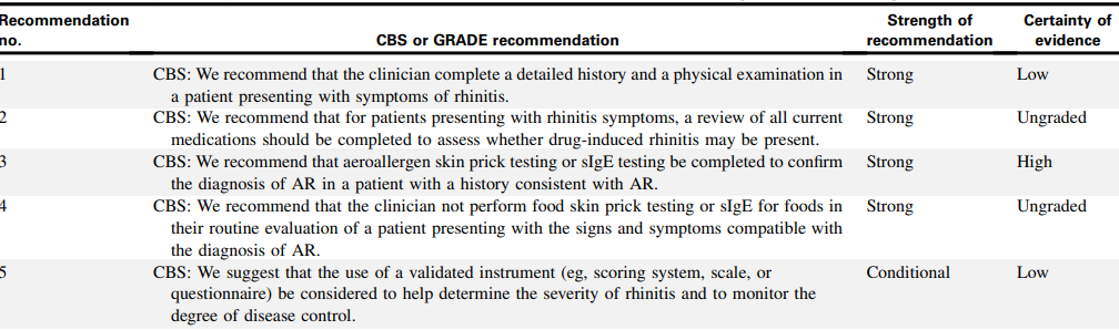
Consensus based statements (CBSs) regarding the diagnosis and management of rhinitis from the JTFPP include the following:51
World Allergy Organization Position Paper
In 2020, the World Allergy Organization published a position paper on IgE allergy diagnostics and other relevant allergy tests. Key statements from the paper can be found below:
- “Clinical suspicion of allergic sensitization is confirmed by demonstrating the presence of allergen-specific IgE antibodies in vivo (skin tests) or in vitro.
- Confirmation of allergen sensitization and the identification of causal allergens are essential for optimizing the management of allergic conditions.
- Skin prick testing (SPT) is the most frequently used method for the detection of IgE antibodies, due to its rapidity, simplicity, and low cost. Skin prick tests and other skin test results must be interpreted by a clinician with adequate knowledge of medical history, clinical findings, and relevant type I allergens (including environmental, food, animal, insect, fungal, and drug allergens). Skin tests should include the relevant allergens in the given geographical area and ideally carried out only using standardized allergenic extracts.
- In vitro tests, including molecular based allergy diagnostics, using either in single-plex and in multi-plexed strategies and other more functional tests, such as Basophil Activation Tests allow to better define the IgE profile of the patient. This approach is in line with the Precision Medicine statements.”57
The paper also states that “Skin tests, especially SPT, represent the most reliable and cost-effective tool for the diagnosis and management of IgE-mediated diseases. They demonstrate a good correlation with outcomes of nasal, conjunctival, dermal, oral, and bronchial challenges.”57
Clinical conditions where SPT is indicated include:
- “Asthma;
- Rhinitis/rhinosinusitis/rhino-conjunctivitis/conjunctivitis;
- Eczema/atopic dermatitis (in the setting of selectively high clinical suspicion for underlying presence of IgE hypersensitivity to specific allergens);
- Suspected food allergy (oral allergy syndrome, anaphylaxis/acute onset or exacerbation of urticaria or eczema that is temporally correlated with food ingestion);
- Suspected drug allergy;
- Hymenoptera venom allergy (systemic reactions immediately following insect sting);
- Suspected occupational disease or exposure to selected potential allergens;
- Chronic urticaria in rare, selected cases which strongly suggest an allergen as potential trigger/ aggravating factor;
- Less common disorders, such as eosinophilic esophagitis, eosinophilic gastroenteritis, or allergic bronchopulmonary a
Skin prick testing “is not routinely indicated in the following instances in the absence of other existing features of allergic disease:
- Suspected food intolerance (e.g., irritable bowel syndrome, etc.);
- Chronic urticaria in the absence of allergic features in the history;
- Desire to lose weight (according to nonconventional approaches, obesity may be due to food intolerance, but no supporting scientific data have been reported in the literature);
- Non-specific food-associated symptoms to food additives/preservatives/colorants;
- Evaluation of the effectiveness of allergen immunotherapy (but may be supportive in Hymenoptera venom immunotherapy);
- Non-specific respiratory symptoms to irritants (i.e., smoke, perfumes, detergents, chemicals and other strong odors);
- Screening for allergic sensitization patterns in the absence of clinical symptoms (i.e., family history of allergy);
- Non-specific cutaneous rashes in the absence of atopic features or other allergic symptoms;
- Migraine, except for the indication of specific hypersensitivity to hormones. However, strong scientific data are still missing;
- Chronic fatigue syndrome.”57
In a 2020 publication on anaphylaxis guidance, the WAO confirms that “allergy testing should be based on patient history and local data regarding the common causes of anaphylaxis in the region. The most frequent elicitor groups worldwide are food, insect venom, and drugs.”58
Choosing Wisely Canada
In 2025, Choosing Wisely published recommendations on allergy and clinical immunology including:
- “Don’t order specific IgG testing to panels of foods. The presence of IgG to a specific food indicates previous exposure not hypersensitivity. The use of methods other than serum-specific IgE evaluation or skin prick testing in diagnosing allergies is not proven and can result in inappropriate diagnosis and treatment.”
- “Don’t order specific immunoglobin E (IgE) tests (skin or serum) unless indicated by the patient’s history specific to that food. Specific IgE to foods may be detectable when the patient is clinically tolerant. Frequent false positives lead to incorrect diagnosis of food allergies and unnecessary dietary restrictions. Appropriate diagnosis and treatment of allergies requires specific IgE testing (either skin or blood tests) based on the patient’s clinical history of signs and symptoms to optimize both cost effectiveness and patient care.”
- “Don’t diagnose or manage current or chronic or persistent asthma in patients >6 years of age without objective testing such as spirometry or a methacholine challenge. Findings on a patient’s history and physical exam such as cough, wheeze and dyspnea may be caused by many conditions, including asthma. When the diagnosis of current or persistent asthma is suspected it must be confirmed with objective testing, as up to one third of patients with suspected asthma show no objective evidence when later tested and may have went into sustained clinical remission or never had asthma. Misdiagnosis leads to delayed treatment of the underlying condition and unnecessary exposure to medication side effects. Objective methods of confirming the diagnosis of asthma in patients in whom asthma is suspected should be used such as spirometry, methacholine challenge, exercise challenge or peak flow variability. These tests may be normal when on treatment.”59
World Allergy Organization (WAO), Allergic Rhinitis and its Impact on Asthma (ARIA), and the Global Allergy and Asthma European Network (GA2LEN)
The WAO, ARIA, and GA2LEN published a consensus document in 2020 focused on molecular based allergy diagnoses. Precision allergy molecular diagnostic applications (PAMD@) “can increase the accuracy of an allergy diagnosis in certain circumstances. In allergic patients, a molecular approach is suitable for the following:
- Assessing the risk of potential allergic reactions, which depend on the individual allergic (clinical) sensitization profile;
- Evaluating whether unknown potential triggering factors are present (i.e., the presence of sIgE versus allergenic molecules correlated with high risk for allergic reactions).”60
National Institute of Allergy and Infectious Diseases (NIAID)
The NIAID convened an expert panel to review current information and to make recommendations related to the evaluation of food allergy (FA), including the use of specific IgE (sIgE) testing.50 With regards to allergen-specific serum IgE determination, NIAID recommended that “sIgE tests for identifying foods that potentially provoke IgE-mediated food-induced allergic reactions, but alone these tests are not diagnostic of FA.” It stated that “sIgE testing and skin prick testing both depend on the presence of allergen-specific antibodies. Because the former test measures sIgE in the serum and the latter reflects IgE bound to cutaneous mast cells, their results may not always correlate. Serum testing can be especially useful when SPTs cannot be done (for example, due to extensive dermatitis or dermatographism), or when antihistamines cannot be discontinued.” The NIAID also recommended not using the combination of SPT, sIgE tests and atopy patch test (ATP) for the routine diagnosis of FA.
Additionally, the NIAID notes that “the routine use of measuring total serum IgE should not be used to make a diagnosis of FA.”50
“Non-standardized tests” such as basophil histamine release/activation, lymphocyte stimulation, allergen-specific IgG, cytotoxicity assays, and mediator release assays should not be used in the routine evaluation of FA, according to the NIAID guidelines.50
In 2017, the NIAID published addendum guidelines for the prevention of peanut allergy in the United States. These guidelines note that the expert panel (EP) “recommends that evaluation with peanut-specific IgE (peanut sIgE) measurement, SPTs, or both be strongly considered before introduction of peanut to determine if peanut should be introduced and, if so, the preferred method of introduction. To minimize a delay in peanut introduction for children who may test negative, testing for peanut sIgE may be the preferred initial approach in certain health care settings, such as family medicine, pediatrics, or dermatology practices, in which skin prick testing is not routine.”61 Further, “The EP does not recommend food allergen panel testing or the addition of sIgE testing for foods other than peanut because of their poor positive predictive value, which could lead to misinterpretation, overdiagnosis of food allergy, and unnecessary dietary restrictions.”61 More, if an infant has severe eczema, an egg allergy, or both, the EP recommends to “Strongly consider evaluation by sIgE measurement and/or SPT and, if necessary, an OFC. Based on test results, introduce peanut-containing foods.”61
American Academy of Pediatrics (AAP)
In 2012, AAP released a clinical report on allergy testing in childhood. It stated that “Both serum sIgE tests and SPT are sensitive and have similar diagnostic properties.” The AAP summary included the following:
- “Treatment decisions for infants and children with allergy should be made on the basis of history and, when appropriate, identified through directed serum sIgE or SPT testing. Newer in vitro sIgE tests have supplanted radioallergosorbent tests.
- Positive sIgE test results indicate sensitization but are not equivalent to clinical allergy. Large panels of indiscriminately performed screening tests may, therefore, provide misleading information.
- Increasingly higher levels of sIgE (higher concentrations on serum tests or SPT wheal size) generally correlate with an increased risk of clinical allergy.
- Use of a multiallergen serum test can be helpful for screening for atopic disease if there is a clinical suspicion. If positive, allergen-specific testing may be considered.
- Tests for allergen-specific IgG antibodies are not helpful for diagnosing allergies.”62
In 2019, the AAP published new guidelines on the prevention of childhood food allergies and other allergic conditions. This article states that “The new recommendations for the prevention of peanut allergy are based largely on the LEAP trial and are endorsed by the AAP.” The AAP endorsed guidelines were published by Togias, et al. (2017) and are noted above. They state that the highest-risk infants (those with severe eczema and/or egg allergies) should be introduced to peanuts by four to six months; further, allergy testing is strongly advised before peanut introduction. SPT and blood testing for peanut-specific IgE (sIgE) are allowable.63,64
In 2020, the AAP published a state-of-the-art review of peanut allergy testing advances and controversies. The article states that “current first-line diagnostic tests for peanut allergy have limited specificity, which may be enhanced with emerging tools such as component-resolved diagnostics.” Like the 2019 guideline, they note that first-line best practices for peanut allergy testing include SPT or serum peanut-specific IgE measurement. While both tests are highly sensitive, neither correlate strongly with reaction severity, according to the AAP.65
U.S. Food and Drug Administration on Xolair
The availability of Xolair for treatment of allergic asthma also has implications for allergy testing. According to the package insert, Xolair is indicated for patients six years of age and older with moderate to severe persistent asthma who have a positive skin test or in-vitro reactivity to a perennial aeroallergen and whose symptoms are inadequately controlled with inhaled corticosteroids… Determine dose (mg) and dosing frequency by serum total IgE level (IU/mL) measured before the start of treatment, and by body weight.51 The prescribing information also notes that “total IgE levels are elevated during treatment and remain elevated for up to one year after the discontinuation of treatment. Therefore, re-testing of IgE levels during Xolair treatment cannot be used as a guide for dose determination.”66
International Consensus Statement on Allergy and Rhinology: Allergic Rhinitis
The authors reviewed the existing evidence behind various aspects of evaluation and diagnosis of the AR patient, and developed the following recommendations for AR diagnostic modalities:67
- Patient history: "Using history to make a presumptive diagnosis of AR is reasonable and would not delay treatment initiation. History should be combined with physical examination, which may not be possible in some scenarios such as telemedicine. Confirmation with diagnostic testing is required for progression to AIT or targeted avoidance therapy, or desirable with inadequate response to treatment… Despite low level evidence specifically addressing this area, history is essential in the diagnosis of AR.”
- Physical examination: “When possible, physical examination should be performed with appropriate personal protective equipment to aid in the diagnosis of AR and exclusion of other conditions. When combined with patient history, it increases diagnostic accuracy and may exclude alternative causes of symptoms.”
- Nasal endoscopy: “Nasal endoscopy may be considered as a diagnostic adjunct in the evaluation of patients with suspected AR.”
- Radiologic studies: “Routine use of imaging is not recommended for the diagnosis of AR.”
- Use of validated subjective instruments and patient reported outcome measures: “Validated surveys may be used to screen for AR, follow treatment outcomes and as a primary outcome measure for clinical trials. Specific tests are optimized for various clinicopathological scenarios.”
- Skin prick testing: “Patients can benefit from identification of their specific sensitivities. Skin prick testing (SPT) is a quick and relatively comfortable way to test several antigens with accuracy similar to other available methods of testing… Regular use of the same SPT device type will allow clinicians to familiarize themselves with it and interpretation of results may therefore be more consistent. The use of standardized allergen extracts can further improve consistency of interpretation.”
- Skin intradermal testing: “Intradermal skin tests may not perform as well as SPT in most clinical situations… Intradermal testing may be used to determine aeroallergen sensitization in individuals suspected of having AR.”
- Blended skin testing techniques: “While AIT can be based off SPT results alone, endpoint-based immunotherapy may have possible benefits of decreased time to therapeutic dosage… Blended skin testing techniques, such as modified quantitative testing, are methods that can be used to determine a starting point for AIT or confirm allergic sensitization.”
- Serum total immunoglobulin E: “Assessment of total IgE may be useful to assess overall atopic status; furthermore, in selected cases it might help guide therapy (i.e., predict outcome of AIT).”
- Serum allergen-specific immunoglobulin E: “Patients can benefit from identification of their specific sensitivities. Further, in some patients who cannot undergo SPT, serum sIgE testing is a safe and effective alternative… Serum sIgE testing may be used in patients who cannot undergo allergy skin testing. The use of highly purified allergen or recombinants can increase the sensitivity, specificity, and diagnostic accuracy of sIgE tests. Rigorous proficiency testing on the part of laboratories may also improve accuracy.”
- Nasal allergen-specific immunoglobulin E: “In patients with non-allergic rhinitis who also have risk factors for atopic disease and have inadequate response to pharmacotherapy, testing for nasal sIgE may be helpful in confirming a diagnosis of local AR and allowing for treatment with AIT. There is no consensus for levels of nasal sIgE that indicate sensitivity… Measurement of nasal sIgE is an option in patients with non-allergic rhinitis suspected of having local AR to support this diagnosis and guide AIT if pharmacologic therapies are inadequate. Consensus for levels of nasal sIgE indicating AR need to be established.”
- Basophil activation test: “The evidence does not support routine use for the diagnosis of AR or for following AIT response… Application of basophil activation test in specific situations where other diagnostic procedures for AR are not possible or conflicting. Potentially useful for monitoring AIT if other methods fail or show conflicting results.”
- Component resolved diagnostic testing: “Molecular diagnosis may be a useful tool for assessment of AR in some scenarios, especially in polysensitized patients… Component resolved diagnostic testing is an option for diagnosis of AR by specialists.”
- Nasal provocation testing: “Application of nasal provocation testing is useful in local AR and to confirm occupational rhinitis.”
- Nasal cytology: “Nasal cytology could help in cases of non-allergic rhinitis to suspect local AR or in cases of AR to diagnose a mixed rhinitis. It could be considered an option in cases of negative SPT and/or serum sIgE to evaluate the presence of mucosal eosinophils and consideration of local AR or type two inflammation. The cut-off values for determining non-allergic rhinitis with eosinophilia syndrome (NARES) are not yet clear.”
- Nasal histology: “Nasal histology may be helpful in clinical research or selected cases (e.g., evaluation of tissue eosinophils during surgery). Recommendation against in routine clinical practice for AR evaluation due to invasive nature of obtaining a specimen.”
- Rhinomanometry: “Rhinomanometry is useful in distinguishing between structural and soft tissue causes of obstruction, when history and examination findings are not congruent, as well as a research tool. Better with individual nasal cavity assessment and four-phase rhinomanometry.”
- Acoustic rhinometry: “Acoustic rhinometry is most useful in research setting as opposed to as a clinical diagnostic tool.”
- Peak nasal inspiratory flow: “Use in conjunction with patient reported outcome measures to improve utility.”
- Nitric oxide measurements: “There is inconsistent evidence in the ability of FeNO or nNO to differentiate adults and children with AR and non-allergic rhinitis. Most studies were of low evidence or small impact. There is no agreed upon cut-off value when performing FeNO or nNO for the diagnosis of AR… History and physical, diagnostic skin testing, or sIgE testing should be the first-line evaluation of AR. FeNO or nasal NO testing may provide additional diagnostic information if necessary but should not be routinely employed for AR diagnosis.”
The National Academies of Science, Engineering and Medicine
The National Academies of Science, Engineering and Medicine convened an expert committee to review the science and management practices of FA. Overall, they found that:
- “Currently, no simple diagnostic tests exist for food allergy.
- Food allergy evaluation procedures include a medical history and physical examination, and also may include food-specific skin prick test, food-specific serum immunoglobulin E test, diagnostic food elimination diet, and oral food challenge (OFC). Selection of the specific tests needs to be individualized based on the medical history of each patient.
- The BAT shows promising preliminary data; the potential utility is recognized and will require additional validation and standardization. Guidelines suggest not using the BAT clinically on the grounds that it is nonstandardized, but recognize its use as a research tool.”68
In 2017, the National Academies of Science, Engineering and Medicine convened an expert committee to examine critical issues related to FA. Regarding diagnosis and prognosis, the committee notes that “physicians [should] use evidence-based, standardized procedures as the basis for FA diagnosis and avoid nonstandardized and unproven procedures…. When food allergy is suspected, the patient should be evaluated by a physician who has the training and experience to select and interpret appropriate diagnostic tests.”69
American Academy of Family Physicians (AAFP)
American Academy of Family Physicians recommendations for practice state: “Allergy and immunologic testing can help clarify the diagnosis and guide treatment. Immediate immunoglobulin E (IgE) and delayed T cell–mediated reactions are the main types of allergic responses. The allergens suspected in an immediate IgE-mediated response are identified through serum IgE-specific antibody or skin testing. For patients with an inhalant allergy, skin or IgE-specific antibody testing is preferred. In patients with food allergies, eliminating the suspected allergenic food from the diet is the initial treatment. If this is ineffective, IgE-specific antibody or skin testing can exclude allergens. An oral food challenge should be performed to confirm the diagnosis. Patients with an anaphylactic reaction to an insect sting should undergo IgE-specific antibody or skin testing. Skin testing for penicillin has a high negative predictive value and can help when penicillin administration is indicated and there are limited alternatives. Testing for other drug allergies has less well-determined sensitivity and specificity but can guide the diagnosis. Patch testing can help identify the allergen responsible for contact dermatitis.”2
European Academy of Allergy and Clinical Immunology (EAACI)
The EAACI published guidelines on “Biomarkers for monitoring the clinical efficacy of allergen immunotherapy (AIT).” In it, they concluded that “to date, there are no validated and generally accepted candidate biomarkers that are predictive or indicative of the clinical response to AIT.” However, they did note sIgE/tIgE ratio and IgE‐FAB as candidate biomarkers for future research.70
The EAACI released a position statement on the BAT. In it, they concluded that “Basophil activation test has been established as a routine diagnostic test with standardized allergen preparations in a number of service laboratories… An important next step is the standardization and automation of analysis of BAT. Once that is achieved, it will be possible to do large multicenter trials to characterize the diagnostic performance of BAT and broaden its use as a clinical tool.”71
The EAACI released a Molecular Allergology User's Guide 2.0. While not a formal guideline, the guide provides comprehensive information about molecular allergen testing. In terms of IgE antibody testing, guide notes that “The clinical relevance of allergen-specific IgE detection in a patient’s serum is strictly as a marker for allergic sensitisation (risk for allergy) and it alone cannot predict the probability of an allergic reaction. The determination of the diagnostic sensitivity and specificity of IgE antibody assays will thus remain difficult to definitively determine because of the lack of an absolute (gold standard) method of defining the presence of allergic disease. This means that the clinical relevance of an allergic sensitisation (i.e. presence of allergen-specific IgE) independent of the use of allergen extracts or molecules for diagnostic purposes will ultimately be determined only by the physician and not by the test.”72
In terms of basophil activation testing, the guide notes that “the BAT can be useful to confirm the diagnosis of food, venom and respiratory allergies.”72
In terms of In vivo testing, the guide notes that “provocation tests are especially helpful when discrepancies exist between the clinical history and other in vivo or in vitro test results, to phenotype patients and to monitor the efficacy of allergen-specific immunotherapy” but further states that “the use of recombinant allergens in provocation tests seems to improve their accuracy; however, it is an unmet need which requires further investigations.”72
Proprietary testing National Institute for Health and Care Excellence (NICE)
National Institute for Health and Care Excellence published a guideline on asthma, recommending against use of serum total or specific IgE for diagnosing asthma. Specific IgE or prick tests to house dust mite should be used to identify triggers to asthma after a formal diagnosis has been made.73
The NICE also released a statement on multiplex allergen testing, particularly “ImmunoCAP ISAC” Although they acknowledge the test’s promise, they state that there is “insufficient evidence to recommend the routine adoption of multiplex allergen testing with ImmunoCAP ISAC 112 to help diagnose allergy and predict the risk of an allergic reaction in people with allergy that is difficult to diagnose, when used with standard clinical assessment.”74
Regarding the assessment and diagnosis of FA in under 19s, NICE published the below recommendations:
For food allergies classified as IgE-mediated:
“Based on the results of the allergy-focused clinical history, if IgE-mediated allergy is suspected, offer the child or young person a skin prick test and/or blood tests for specific IgE antibodies to the suspected foods and likely co-allergens.”
“Tests should only be undertaken by healthcare professionals with the appropriate competencies to select, perform and interpret them.”
“Skin prick tests should only be undertaken where there are facilities to deal with an anaphylactic reaction.”
“Choose between a skin prick test and a specific IgE antibody blood test based on:
- the results of the allergy-focused clinical history and
- whether the test is suitable for, safe for and acceptable to the child or young person (or their parent or carer) and
- the available competencies of the healthcare professional to undertake the test and interpret the results.”
“Do not carry out allergy testing without first taking an allergy-focused clinical history. Interpret the results of tests in the context of information from the allergy-focused clinical history.”
“Do not use atopy patch testing or oral food challenges to diagnose IgE-mediated food allergy in primary care or community settings.”75
The Royal College of Pathologists
The Royal College of Pathologists, based in the United Kingdom, published guidelines for allergy testing in primary care. The guidelines state four key clinical features; “Prior tolerance does not exclude allergy. Symptoms occur within minutes to an hour of exposure to the trigger. The symptoms are reproducible and occur with every subsequent exposure. The symptoms do not occur without exposure to the allergen.”76
For food allergy, the guidelines recommend “only test ASIgE [allergen specific IgE] if the answer is yes to all questions, and limit testing to the relevant trigger food.” The questions referenced are: “Are the Symptoms consistent with an IgE mediated mechanism? Is the Timing consistent with an IgE mediated mechanism? Can symptoms be attributed to a likely Allergen? Are the symptoms Reproducible?”76
For inhalant allergy, the guidelines recommend “ASIgE testing is only advised in primary care if management will be directly affected by the result (e.g. testing for dog dander leading to avoidance) or if testing is required prior to referral (e.g. referral is to ear, nose and throat (ENT) services if results are negative and to allergy or immunology if positive).”76
For bee and wasp venom allergy, the guidelines recommend “Do not test ASIgE in the context of large local reactions or mild local reactions to wasp or bee stings. In the context of systemic reactions to wasp or bee venom, patients should be referred to allergy or immunology services. Blood tests for baseline mast cell tryptase and ASIgE to wasp and bee venom can be sent for analysis at the time of referral.”76
The guidelines also state that “Isolated randomly occurring urticaria with or without angioedema is extremely common. Most cases of urticaria are not caused by allergy but are spontaneous in onset. The lack of a clear and consistent trigger excludes allergy on clinical grounds in these cases and allergy testing is not indicated.”76
References
1. Hamilton R. Overview of Allergens and Environmental Sampling. Updated November 06, 2025. https://www.uptodate.com/contents/overview-of-allergens-and-environmental-sampling
2. Chang, Guarderas. Allergy Testing: Common Questions and Answers. American family physician. Jul 1 2018;98(1):34-39.
3. Kowal K, DuBuske L. Overview of skin testing for IgE-mediated allergic disease. Updated November 03, 2025. https://www.uptodate.com/contents/overview-of-skin-testing-for-allergic-disease
4. Bernstein IL, Li JT, Bernstein DI, et al. Allergy diagnostic testing: an updated practice parameter. Annals of allergy, asthma & immunology : official publication of the American College of Allergy, Asthma, & Immunology. Mar 2008;100(3 Suppl 3):S1-148. doi:10.1016/s1081-1206(10)60305-5
5. CDC. Allergies. Updated January 9, 2026. https://www.cdc.gov/nchs/fastats/allergies.htm
6. Pawankar R, Holgate ST, Canonica GW, Lockey RF, Blaiss MS. WAO White Book on Allergy | World Allergy Organization. 2013.
7. Adams PF, Kirzinger WK, Martinez M. Summary health statistics for the U.S. population: National Health Interview Survey, 2012. Vital and health statistics Series 10, Data from the National Health Survey. Dec 2013;(259):1-95.
8. Zug KA, Pham AK, Belsito DV, et al. Patch testing in children from 2005 to 2012: results from the North American contact dermatitis group. Dermatitis : contact, atopic, occupational, drug. Nov-Dec 2014;25(6):345-55. doi:10.1097/der.0000000000000083
9. Sampson HA, Aceves S, Bock SA, et al. Food allergy: a practice parameter update-2014. The Journal of allergy and clinical immunology. Nov 2014;134(5):1016-25.e43. doi:10.1016/j.jaci.2014.05.013
10. Chow AW, Benninger MS, Brook I, et al. IDSA clinical practice guideline for acute bacterial rhinosinusitis in children and adults. Clinical infectious diseases : an official publication of the Infectious Diseases Society of America. Apr 2012;54(8):e72-e112. doi:10.1093/cid/cir1043
11. Mayo Clinic. Aspergillosis. Updated September 22, 2025. https://www.mayoclinic.org/diseases-conditions/aspergillosis/symptoms-causes/syc-20369619
12. Stokes J, Casale T. The relationship between IgE and allergic disease. Updated October 21, 2024. https://www.uptodate.com/contents/the-relationship-between-ige-and-allergic-disease
13. Kowal K, DuBuske L. Overview of in vitro allergy tests. Updated January 23, 2025. https://www.uptodate.com/contents/overview-of-in-vitro-allergy-tests
14. Rietschel RL. COMPARISON OF ALLERGIC AND IRRITANT CONTACT DERMATITIS. Immunology and Allergy Clinics. 1997/08/01 1997;17(3):359-364. doi:10.1016/S0889-8561(05)70314-3
15. Mowad CM. Patch testing: pitfalls and performance. Current opinion in allergy and clinical immunology. Oct 2006;6(5):340-4. doi:10.1097/01.all.0000244794.03239.8e
16. Kim SY, Kim JH, Jang YS, et al. The Basophil Activation Test Is Safe and Useful for Confirming Drug-Induced Anaphylaxis. Allergy, asthma & immunology research. Nov 2016;8(6):541-4. doi:10.4168/aair.2016.8.6.541
17. Santos AF, Lack G. Basophil activation test: food challenge in a test tube or specialist research tool? Clinical and translational allergy. 2016;6:10. doi:10.1186/s13601-016-0098-7
18. Bahri R, Custovic A, Korosec P, et al. Mast cell activation test in the diagnosis of allergic disease and anaphylaxis. The Journal of allergy and clinical immunology. 2018;142(2):485-496 e16. doi:10.1016/j.jaci.2018.01.043
19. Santos AF, Shreffler WG. Road map for the clinical application of the basophil activation test in food allergy. Clinical and experimental allergy : journal of the British Society for Allergy and Clinical Immunology. Sep 2017;47(9):1115-1124. doi:10.1111/cea.12964
20. Depince-Berger AE, Sidi-Yahya K, Jeraiby M, Lambert C. Basophil activation test: Implementation and standardization between systems and between instruments. Cytometry Part A : the journal of the International Society for Analytical Cytology. Mar 2017;91(3):261-269. doi:10.1002/cyto.a.23078
21. Hemmings O, Kwok M, McKendry R, Santos AF. Basophil Activation Test: Old and New Applications in Allergy. Current Allergy and Asthma Reports. 2018/11/15 2018;18(12):77. doi:10.1007/s11882-018-0831-5
22. Cell Science Systems. Identify food and chemical sensitivities with the Alcat Test. https://cellsciencesystems.com/providers/alcat-test/
23. Beyer K, Teuber SS. Food allergy diagnostics: scientific and unproven procedures. Current opinion in allergy and clinical immunology. Jun 2005;5(3):261-6. doi:10.1097/01.all.0000168792.27948.f9
24. Wuthrich B. Unproven techniques in allergy diagnosis. Journal of investigational allergology & clinical immunology. 2005;15(2):86-90.
25. Hammond C, Lieberman JA. Unproven Diagnostic Tests for Food Allergy. Immunology and allergy clinics of North America. Feb 2018;38(1):153-163. doi:10.1016/j.iac.2017.09.011
26. Genova. Allergix® IgG4 Food Antibodies 90 - Serum. https://www.gdx.net/product/allergix-igg4-food-antibodies-90-food-sensitivity-test-serum
27. Spiriplex. Allergies. https://spiriplex.com/allergies/
28. BioSpace. AllerGenis Allergy Diagnostic Company to Present Significant Milestones at the Biotech Showcase(TM) 2021. https://www.biospace.com/article/allergenis-allergy-diagnostic-company-to-present-significant-milestones-at-the-biotech-showcase-tm-2021/
29. Nelson HS. Variables in Allergy Skin Testing. Immunology and Allergy Clinics. 2001/05/01 2001;21(2):281-290. doi:10.1016/S0889-8561(05)70206-X
30. Werther RL, Choo S, Lee KJ, Poole D, Allen KJ, Tang ML. Variability in Skin Prick Test Results Performed by Multiple Operators Depends on the Device Used. World Allergy Organ J. 2012;5(12):200-4. doi:10.1097/WOX.0b013e31827e6513
31. Oppenheimer J, Nelson HS. Skin testing: a survey of allergists. Annals of allergy, asthma & immunology : official publication of the American College of Allergy, Asthma, & Immunology. Jan 2006;96(1):19-23. doi:10.1016/s1081-1206(10)61034-4
32. Hamilton RG, Matsson PN, Hovanec-Burns DL, et al. Analytical Performance Characteristics, Quality Assurance and Clinical Utility of Immunological Assays for Human IgE Antibodies of Defined Allergen Specificities.(CLSI-ILA20-A3). Journal of Allergy and Clinical Immunology. 2015;135(2):AB8. doi:10.1016/j.jaci.2014.12.961
33. Knight V, Wolf ML, Trikha A, Curran-Everett D, Hiserote M, Harbeck RJ. A comparison of specific IgE and skin prick test results to common environmental allergens using the HYTEC™ 288. Journal of Immunological Methods. 2018/11/01/ 2018;462:9-12. doi:10.1016/j.jim.2018.07.005
34. Carlsson M, Thorell L, Sjolander A, Larsson-Faria S. Variability of total and free IgE levels and IgE receptor expression in allergic subjects in and out of pollen season. Scandinavian journal of immunology. Apr 2015;81(4):240-8. doi:10.1111/sji.12270
35. Siroux V, Lupinek C, Resch Y, et al. Specific IgE and IgG measured by the MeDALL allergen-chip depend on allergen and route of exposure: The EGEA study. The Journal of allergy and clinical immunology. Feb 2017;139(2):643-654.e6. doi:10.1016/j.jaci.2016.05.023
36. Sookrung N, Jotikaprasardhna P, Bunnag C, Chaicumpa W, Tungtrongchitr A. Concordance of skin prick test and serum-specific IgE to locally produced component-resolved diagnostics for cockroach allergy. Annals of allergy, asthma & immunology : official publication of the American College of Allergy, Asthma, & Immunology. Jan 2019;122(1):93-98. doi:10.1016/j.anai.2018.09.463
37. He YT, Reisacher WR. Sensitivity, specificity, and predictive value of oral mucosal brush biopsy for the diagnosis of peanut allergy. Int Forum Allergy Rhinol. Jun 2019;9(6):624-628. doi:10.1002/alr.22302
38. Tschopp JM, Sistek D, Schindler C, et al. Current allergic asthma and rhinitis: diagnostic efficiency of three commonly used atopic markers (IgE, skin prick tests, and Phadiatop). Results from 8329 randomized adults from the SAPALDIA Study. Swiss Study on Air Pollution and Lung Diseases in Adults. Allergy. Jun 1998;53(6):608-13. doi:10.1111/j.1398-9995.1998.tb03937.x
39. Usmani N, Wilkinson SM. Allergic skin disease: investigation of both immediate- and delayed-type hypersensitivity is essential. Clinical and experimental allergy : journal of the British Society for Allergy and Clinical Immunology. Oct 2007;37(10):1541-6. doi:10.1111/j.1365-2222.2007.02805.x
40. Soares-Weiser K, Takwoingi Y, Panesar SS, et al. The diagnosis of food allergy: a systematic review and meta-analysis. Allergy. Jan 2014;69(1):76-86. doi:10.1111/all.12333
41. Klemans RJ, van Os-Medendorp H, Blankestijn M, Bruijnzeel-Koomen CA, Knol EF, Knulst AC. Diagnostic accuracy of specific IgE to components in diagnosing peanut allergy: a systematic review. Clinical and experimental allergy : journal of the British Society for Allergy and Clinical Immunology. Apr 2015;45(4):720-30. doi:10.1111/cea.12412
42. Caglayan Sozmen S, Povesi Dascola C, Gioia E, Mastrorilli C, Rizzuti L, Caffarelli C. Diagnostic accuracy of patch test in children with food allergy. Pediatric allergy and immunology : official publication of the European Society of Pediatric Allergy and Immunology. Aug 2015;26(5):416-22. doi:10.1111/pai.12377
43. Santos AF, Douiri A, Becares N, et al. Basophil activation test discriminates between allergy and tolerance in peanut-sensitized children. The Journal of allergy and clinical immunology. Sep 2014;134(3):645-52. doi:10.1016/j.jaci.2014.04.039
44. Santos AF, Du Toit G, Douiri A, et al. Distinct parameters of the basophil activation test reflect the severity and threshold of allergic reactions to peanut. The Journal of allergy and clinical immunology. Jan 2015;135(1):179-86. doi:10.1016/j.jaci.2014.09.001
45. Davila I, Valero A, Entrenas LM, Valveny N, Herraez L. Relationship between serum total IgE and disease severity in patients with allergic asthma in Spain. Journal of investigational allergology & clinical immunology. 2015;25(2):120-7.
46. Tannert LK, Mortz CG, Skov PS, Bindslev-Jensen C. Positive Skin Test or Specific IgE to Penicillin Does Not Reliably Predict Penicillin Allergy. The journal of allergy and clinical immunology In practice. May - Jun 2017;5(3):676-683. doi:10.1016/j.jaip.2017.03.014
47. Suárez-Fariñas M, Suprun M, Kearney P, et al. Accurate and reproducible diagnosis of peanut allergy using epitope mapping. Allergy. 2021;n/a(n/a)doi:10.1111/all.14905
48. Kwong KY, Chen Z, Scott L, Hilborne LH. Optimizing Identification of Allergic Sensitization to Seasonal Inhalant Allergens in the USA: Implications for Constructing Optimal Panels to Evaluate Patients with Allergy. International archives of alleregy and immunology. 2024;doi:10.1159/000538420
49. AAAAI. Allergy Testing. Updated November 13, 2023. https://www.aaaai.org/tools-for-the-public/conditions-library/allergies/allergy-testing
50. Boyce JA, Assa'ad A, Burks AW, et al. Guidelines for the Diagnosis and Management of Food Allergy in the United States: Summary of the NIAID-Sponsored Expert Panel Report. The Journal of allergy and clinical immunology. Dec 2010;126(6):1105-18. doi:10.1016/j.jaci.2010.10.008
51. Dykewicz MS, Wallace DV, Amrol DJ, et al. Rhinitis 2020: A practice parameter update. The Journal of allergy and clinical immunology. Oct 2020;146(4):721-767. doi:10.1016/j.jaci.2020.07.007
52. Lieberman P, Nicklas RA, Randolph C, et al. Anaphylaxis--a practice parameter update 2015. Annals of allergy, asthma & immunology : official publication of the American College of Allergy, Asthma, & Immunology. Nov 2015;115(5):341-84. doi:10.1016/j.anai.2015.07.019
53. Golden DBK, Wang J, Waserman S, et al. Anaphylaxis: A 2023 practice parameter update. Annals of Allergy, Asthma & Immunology. 2024;132(2):124-176. doi:10.1016/j.anai.2023.09.015
54. AAAAI. Some Tests and Procedures Are Over or Misused to Diagnose and Treat Allergies, Asthma and Immunologic Disorders. Updated April 4, 2012. https://www.aaaai.org/Aaaai/media/MediaLibrary/PDF%20Documents/Media/Choosing-WiselyT.pdf
55. Greenhawt M, Shaker M, Wang J, et al. Peanut allergy diagnosis: A 2020 practice parameter update, systematic review, and GRADE analysis. The Journal of allergy and clinical immunology. Dec 2020;146(6):1302-1334. doi:10.1016/j.jaci.2020.07.031
56. Fonacier L, Bernstein DI, Pacheco K, et al. Contact dermatitis: a practice parameter-update 2015. The journal of allergy and clinical immunology In practice. May-Jun 2015;3(3 Suppl):S1-39. doi:10.1016/j.jaip.2015.02.009
57. Ansotegui, Melioli G, Canonica GW, et al. IgE allergy diagnostics and other relevant tests in allergy, a World Allergy Organization position paper. World Allergy Organ J. Feb 2020;13(2):100080. doi:10.1016/j.waojou.2019.100080
58. Cardona V, Ansotegui IJ, Ebisawa M, et al. World allergy organization anaphylaxis guidance 2020. World Allergy Organ J. Oct 2020;13(10):100472. doi:10.1016/j.waojou.2020.100472
59. Choosing Wisely Canada. Allergy & Clinical Immunology. https://choosingwiselycanada.org/recommendation/allergy-clinical-immunology/
60. Ansotegui, Melioli G, Canonica GW, et al. A WAO - ARIA - GA(2)LEN consensus document on molecular-based allergy diagnosis (PAMD@): Update 2020. World Allergy Organ J. Feb 2020;13(2):100091. doi:10.1016/j.waojou.2019.100091
61. Togias A, Cooper SF, Acebal ML, et al. Addendum guidelines for the prevention of peanut allergy in the United States: Report of the National Institute of Allergy and Infectious Diseases-sponsored expert panel. The Journal of allergy and clinical immunology. Jan 2017;139(1):29-44. doi:10.1016/j.jaci.2016.10.010
62. AAP. Allergy Testing in Childhood: Using Allergen-Specific IgE Tests. https://pdfs.semanticscholar.org/51a4/dfa0a84e4dc8b0893529a6197bd9b94bdbfe.pdf
63. Greer FR, Sicherer SH, Burks AW. The Effects of Early Nutritional Interventions on the Development of Atopic Disease in Infants and Children: The Role of Maternal Dietary Restriction, Breastfeeding, Hydrolyzed Formulas, and Timing of Introduction of Allergenic Complementary Foods. Pediatrics. Apr 2019;143(4)doi:10.1542/peds.2019-0281
64. Sicherer S. New guidelines detail use of ‘infant-safe’ peanut to prevent allergy. https://www.aappublications.org/news/2017/01/05/PeanutAllergy010517
65. Abrams EM, Chan ES, Sicherer S. Peanut Allergy: New Advances and Ongoing Controversies. Pediatrics. 2020;145(5):e20192102. doi:10.1542/peds.2019-2102
66. FDA. Xolair Label. https://www.accessdata.fda.gov/drugsatfda_docs/label/2016/103976s5225lbl.pdf
67. Wise SK, Damask C, Roland LT, et al. International consensus statement on allergy and rhinology: Allergic rhinitis - 2023. Int Forum Allergy Rhinol. Apr 2023;13(4):293-859. doi:10.1002/alr.23090
68. NASEM. Finding a Path to Safety in Food Allergy: Assessment of the Global Burden, Causes, Prevention, Management, and Public Policy. Finding a Path to Safety in Food Allergy: Assessment of the Global Burden, Causes, Prevention, Management, and Public Policy. 2017;doi:10.17226/23658
69. Sicherer SH, Allen K, Lack G, Taylor SL, Donovan SM, Oria M. Critical Issues in Food Allergy: A National Academies Consensus Report. Pediatrics. Aug 2017;140(2)doi:10.1542/peds.2017-0194
70. Shamji MH, Kappen JH, Akdis M, et al. Biomarkers for monitoring clinical efficacy of allergen immunotherapy for allergic rhinoconjunctivitis and allergic asthma: an EAACI Position Paper. Allergy. 2017/08/01 2017;72(8):1156-1173. doi:10.1111/all.13138
71. Hoffmann HJ, Santos AF, Mayorga C, et al. The clinical utility of basophil activation testing in diagnosis and monitoring of allergic disease. Allergy. 2015/11/01 2015;70(11):1393-1405. doi:10.1111/all.12698
72. Dramburg S, Hilger C, Santos AF, et al. EAACI Molecular Allergology User's Guide 2.0. Pediatric allergy and immunology : official publication of the European Society of Pediatric Allergy and Immunology. Mar 2023;34 Suppl 28:e13854. doi:10.1111/pai.13854
73. NICE. Asthma: diagnosis, monitoring and chronic asthma management. Updated November 27, 2024. https://www.nice.org.uk/guidance/ng245
74. NICE. ImmunoCAP ISAC 112 for multiplex allergen testing. Updated May 18, 2016. https://www.nice.org.uk/guidance/dg24/chapter/1-Recommendations
75. NICE. Food allergy in under 19s: assessment and diagnosis. National Institute for Health and Care Excellence (NICE). Updated February 23, 2011. https://www.nice.org.uk/guidance/cg116/chapter/Recommendations#ige-mediated-food-allergy
76. The Royal College of Pathologists. Guidelines for the use of laboratory allergy testing in primary care. https://www.rcpath.org/static/a49f66cc-3c78-4ee7-97814c34a9d7d462/G194-BPR-Use-of-laboratory-allergy-testing-in-primary-care.pdf
Coding Section
| Code |
Number |
Code Description |
| CPT |
82784 |
Gammaglobulin (immunoglobulin); IgA, IgD, IgG, IgM, each |
|
|
82785 |
Gammaglobulin; IgE |
|
|
82787 |
Immunoglobulin subclasses (e.g.: IgG 1, 2, 3, or 4) each |
|
|
83516 |
Immunoassay for analyte other than infectious agent antibody or infectious agent antigen; qualitative or semiquantitative, multiple step method (ALCAT) |
|
|
86001 |
Allergen specific IgG quantitative or semi-quantitative, each allergen |
|
|
86003 |
Allergen specific IgE quantitative or semi-quantitative, each allergen |
|
|
86005 |
Qualitative, multi-allergen screen (dipstick, paddle or disk) |
|
|
86008 |
Allergen specific IgE; quantitative or semiquantitative, recombinant or purified component, each |
|
|
88184 |
Flow cytometry, cell surface, cytoplasmic, or nuclear marker, technical component only; first marker [anti-IgE receptor antibody testing](BAT) |
|
|
88185 |
Flow cytometry, cell surface, cytoplasmic, or nuclear marker, technical component only; each additional marker |
| 95004 | Percutaneous tests (scratch, puncture, prick) with allergenic extracts, immediate type reaction, including test interpretation and report, specify number of tests | |
|
|
0165U |
Peanut allergen-specific quantitative assessment of multiple epitopes using enzyme-linked immunosorbent assay (ELISA), blood, individual epitope results and probability of peanut allergy |
|
|
0178U |
Peanut allergen-specific quantitative assessment of multiple epitopes using enzyme-linked immunosorbent assay (ELISA), blood, report of minimum eliciting exposure for a clinical reaction |
| ICD-10 Codes |
F78.A1 |
SYNGAP1-related intellectual disability |
|
|
F78.A9 |
Other genetic related intellectual disability |
|
|
R05.1 |
Acute cough |
|
|
R05.2 |
Subacute cough |
|
|
R05.3 |
Chronic cough |
|
|
R05.4 |
Cough syncope |
|
|
R05.8 |
Other specified cough |
|
|
R05.9 |
Cough, unspecified |
|
|
Z91.010 |
Allergy to peanuts |
|
|
Z91.011. |
Allergy to milk products |
|
|
Z91.012 |
Allergy to eggs |
|
|
Z91.013 |
Allergy to seafood |
|
|
Z91.018 |
Allergy to other foods |
|
|
Z91.02 |
Food additives allergy status |
|
|
Z91.030 |
Bee allergy status |
|
|
Z91.038 |
Other insect allergy status |
|
|
Z91.040 |
Latex allergy status |
|
|
Z91.041 |
Radiographic dye allergy status |
|
|
Z91.048 |
Other nonmedicinal substance allergy status |
|
|
Z91.09 |
Other allergy status, other than to drugs and biological substances |
|
|
Z91.14 |
Patient's other noncompliance with medication regimen |
Procedure and diagnosis codes on Medical Policy documents are included only as a general reference tool for each policy. They may not be all-inclusive.
This medical policy was developed through consideration of peer-reviewed medical literature generally recognized by the relevant medical community, U.S. FDA approval status, nationally accepted standards of medical practice and accepted standards of medical practice in this community and other nonaffiliated technology evaluation centers, reference to federal regulations, other plan medical policies, and accredited national guidelines.
"Current Procedural Terminology © American Medical Association. All Rights Reserved"
History From 2013 Forward
| 05/07/2026 | Annual review, adding statement on IgE in-vitro testing when performed on same day as skin prick testing. Also updating table of terminology, rationale, and references. Adding CPT code 95004 |
| 04/14/2025 | Annual review, no change to policy intent. Updating description, coverage criteria 1.h removing “and PIDs involving T, NK”, rationale and references. |
| 04/22/2024 | Annual review, no change to policy intent. Policy is be updated for clarity and consistency. Coverage criteria 7 and 8 are being merged into one statement, Also updating description, table of terminology, rationale and references. |
| 04/01/2024 | Annual review, no change to policy intent. |
| 04/18/2023 | Interim review, annual review now moved to April. NO change to policy intent, but, policy is being rewritten for clarity and consistency. Also updating description, table of terminology, rationale and references. |
| 01/05/2023 | Annual review, no change to policy intent. |
| 04/27/2022 |
Interim review. Adding coverage criteria #4 and updating criteria 1a. Also updating rationale and references. |
| 10/01/2021 |
Annual review, adding criteria #9 related to non-specific IgE testing. Also updating background, rationale, references and regulatory status. |
| 10/01/2020 |
Annual update, Updating rationale, references, description, policy and coding to include verbiage related to peanut allergy testing. |
| 01/10/2020 |
Interim review, updating criteria for RAST, MAST, FAST, ELISA and ImmunoCAP testing. NO other changes made. |
| 10/23/2019 |
Annual review, Updating Title, description, policy, rationale, regulatory status, references and coding. No other changes made.. |
| 10/09/2019 |
Annual review, no change to policy intent. |
| 11/27/2018 |
Interim review adding investigational verbiage for basophil activation flow cytometry. No other changes. |
| 10/01/2018 |
Annual review, no change to policy intent. |
| 12/7/2017 |
Updating policy with 2018 coding. No other changes. |
| 10/30/2017 |
Interim review adding the following policy verbiage:. No other changes made. |
| 10/02/2017 |
Annual review, no change to policy intent. Updated coding with 2018 codes. |
| 06/19/2017 |
Updated coding section. No other changes. |
| 04/25/2017 |
Updated category to Laboratory. No other changes. |
| 11/07/2016 |
Interim review to place correct number of allergen specific antibodies from 36 to 20. |
| 09/21/2016 |
Annual review, Updating to add coding and verbiage to policy that states" In-vitro specific IgE testing is limited to 20 allergen specific antibodies per year. Additional testing beyond this number will require individual review for coverage criteria. |
| 10/07/2014 |
Annual review, no changes made. |
| 10/30/2013 |
Added Related Policy Section. |